María Jimena Costa
Ph.D. in Computer Science - Medical Image Processing
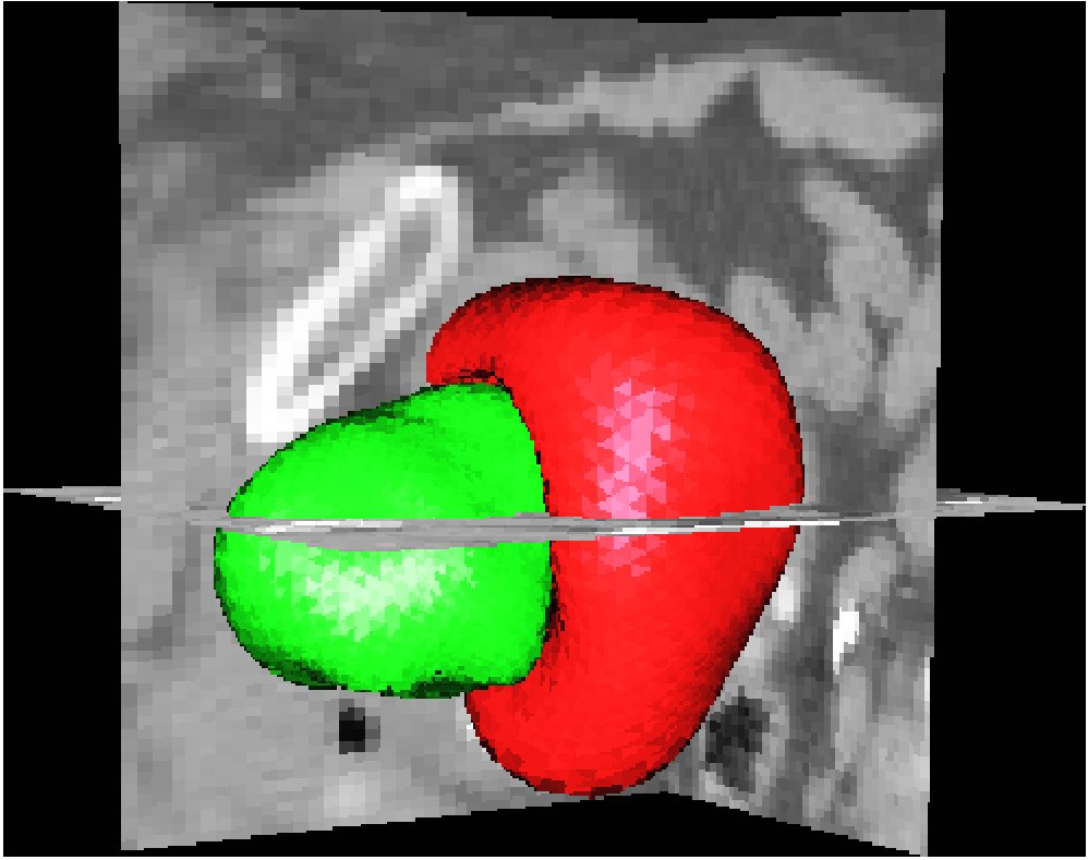
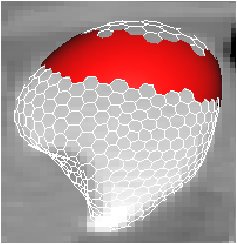
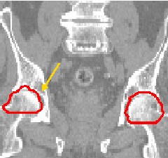
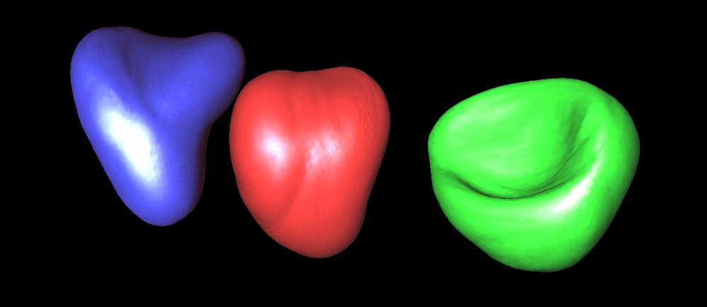
Automatic Segmentation of Bladder and Prostate Using Coupled 3D Deformable Models.
Costa J., Delingette H. and Ayache N.
ISBI 2007
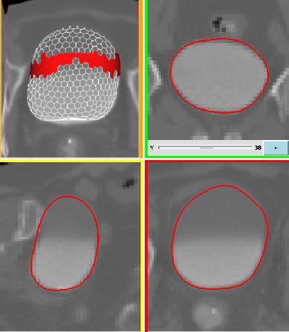
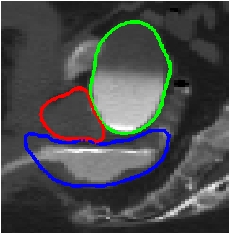
We are interested in the fully automatic delineation of the bladder in CT images in the frame of dose calculation for conformational radiotherapy. To this end we fit a series of 3D deformable templates to the contours of anatomical structures. The novelty of our approach resides in the ability to automatically adapt to different kinds of bladder images (homogenous, non--homogenous, contrasted or non--contrasted). The adaptation of the algorithm to inhomogeneities within the bladder improves the accuracy of the segmentation. We validate our approach on a database of tomodensitometric (CT) images of the lower abdomen of male patients.
Publications
Segmentation of Anatomical Structures of the Lower Abdomen using 3D Deformable Surfaces.
Costa, J.Ph.D. Thesis, March 2008
An Efficient Locally Affine Framework for the Smooth Registration of Anatomical Structures.
Commowick O., Arsigny V., Isambert A., Costa J., et al.Medical Image Analysis 2008
Automatic Segmentation of Bladder and Prostate Using Coupled 3D Deformable Models.
Costa J., et al.MICCAI 2007
Automatic Segmentation of the Bladder Using Deformable Models.
Costa, J., et al.ISBI 2007
An Efficient Locally Affine Framework for the Registration of Anatomical Structures.
Commowick O., Arsigny V., Costa J., Ayache N. and Malandain G.ISBI 2006
Towards an automatic delineation of lower abdomen structures for conformational radiotherapy based on CT images.
Costa J., et al.SFPM 2005.

Distributed Ontological Encoding Through Symbol Recirculation.
Costa J., Duboue P.ASAI 2004.